Carotid plaque on ultrasound: what it means and what to do next
A carotid plaque on a neck ultrasound does not mean a stroke is inevitable. How it differs from intima-media thickening, why stenosis matters and how often the scan is repeated.
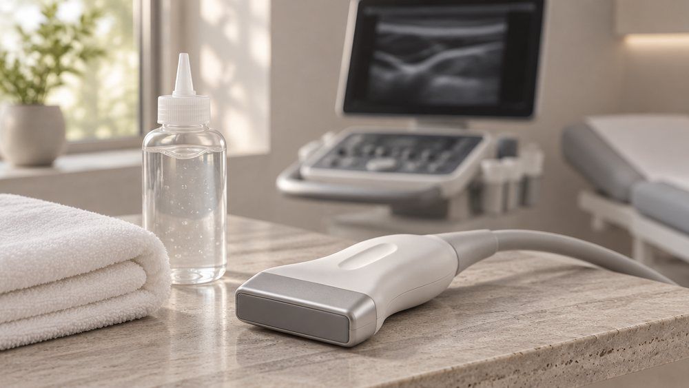
A carotid plaque found on a neck ultrasound does not mean a stroke is inevitable. It shows that atherosclerosis is already present, and the next step is to assess how advanced it is. What happens next depends on the size of the plaque, how much it narrows the artery, its characteristics and your overall cardiovascular risk.
“Doctor, they found a plaque in my carotid artery. Is it dangerous? Could I have a stroke?” This is a common reaction to the result, especially for people who until then considered themselves perfectly healthy.
What is a carotid plaque?
A carotid plaque is a build-up of lipids and other components in the wall of a carotid artery. It forms through atherosclerosis, a chronic process that develops inside the artery wall.
The carotid arteries supply blood to the brain, and they can be examined with ultrasound. During a carotid scan, the doctor assesses the vessel walls, any plaques, how much the lumen is narrowed and the speed of blood flow.
Is a plaque the same as increased intima-media thickness?
No, these are two different findings. Carotid intima-media thickness (CIMT) measures the inner layers of the artery wall, and an increase does not always mean there is an atherosclerotic plaque. A plaque is a localised atherosclerotic change in the vessel wall that protrudes into the artery’s lumen.
Can a small carotid plaque be ignored?
A small plaque that does not disturb blood flow usually does not need surgery, but it should not simply be dismissed. Its presence can point to a higher long-term cardiovascular risk.
That is why the doctor looks beyond the neck arteries and also assesses other risk factors:
- LDL, or “bad” cholesterol;
- ApoB, when needed;
- lipoprotein(a), or Lp(a);
- blood pressure;
- diabetes;
- smoking;
- family history.
What matters more: plaque size or the degree of stenosis?
Both matter. On a carotid ultrasound, the doctor determines:
- where the plaque sits and how large it is;
- how much the artery is narrowed (the degree of stenosis);
- the plaque’s characteristics, including signs of possible instability;
- blood flow velocities.
Significant carotid stenosis calls for particular attention, especially in someone who has already had a transient ischaemic attack (TIA) or a stroke.
Can medication remove a plaque?
Medication usually cannot remove a plaque completely: the main goal of treatment is to lower the chance that atherosclerosis progresses and leads to cardiovascular complications. This involves addressing risk factors and, where clinically indicated, lipid-lowering therapy, primarily statins.
With intensive LDL lowering, some plaques can stabilise, and partial regression — a modest reduction in plaque size — is sometimes seen. Expecting the plaque to vanish entirely, however, is unrealistic.
One more important point. A small plaque without symptoms is not, on its own, a reason to start antiplatelet medication — the tablets that reduce platelet clumping and are often called “blood thinners”.
How often should a carotid ultrasound be repeated?
There is no single interval that suits everyone: it depends on the degree of stenosis, the plaque’s characteristics, symptoms and overall risk. With a small plaque and no significant stenosis, frequent repeat scans are usually unnecessary, and the scan is typically done once a year. With significant stenosis, the doctor sets an individual follow-up schedule.
Who needs a carotid Doppler scan?
A carotid ultrasound is not a mandatory annual screening test for every healthy adult. It is, however, usually recommended for people with raised LDL cholesterol.
The bottom line: a carotid plaque is not a verdict, but it is also not an incidental finding that can always be ignored. It offers a chance to detect atherosclerosis before serious complications develop and to start prevention in good time.
At Longevium in Dubai, a carotid Doppler ultrasound is part of Cholesterol Check 30+, together with an extended lipid panel — LDL, ApoB and Lp(a) — and a consultation with a cardiologist-lipidologist.